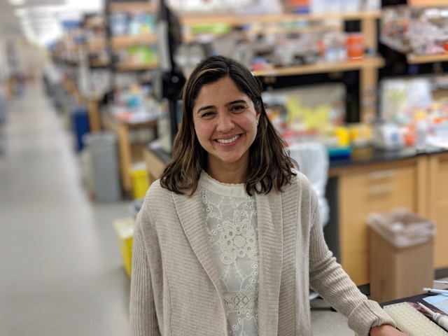
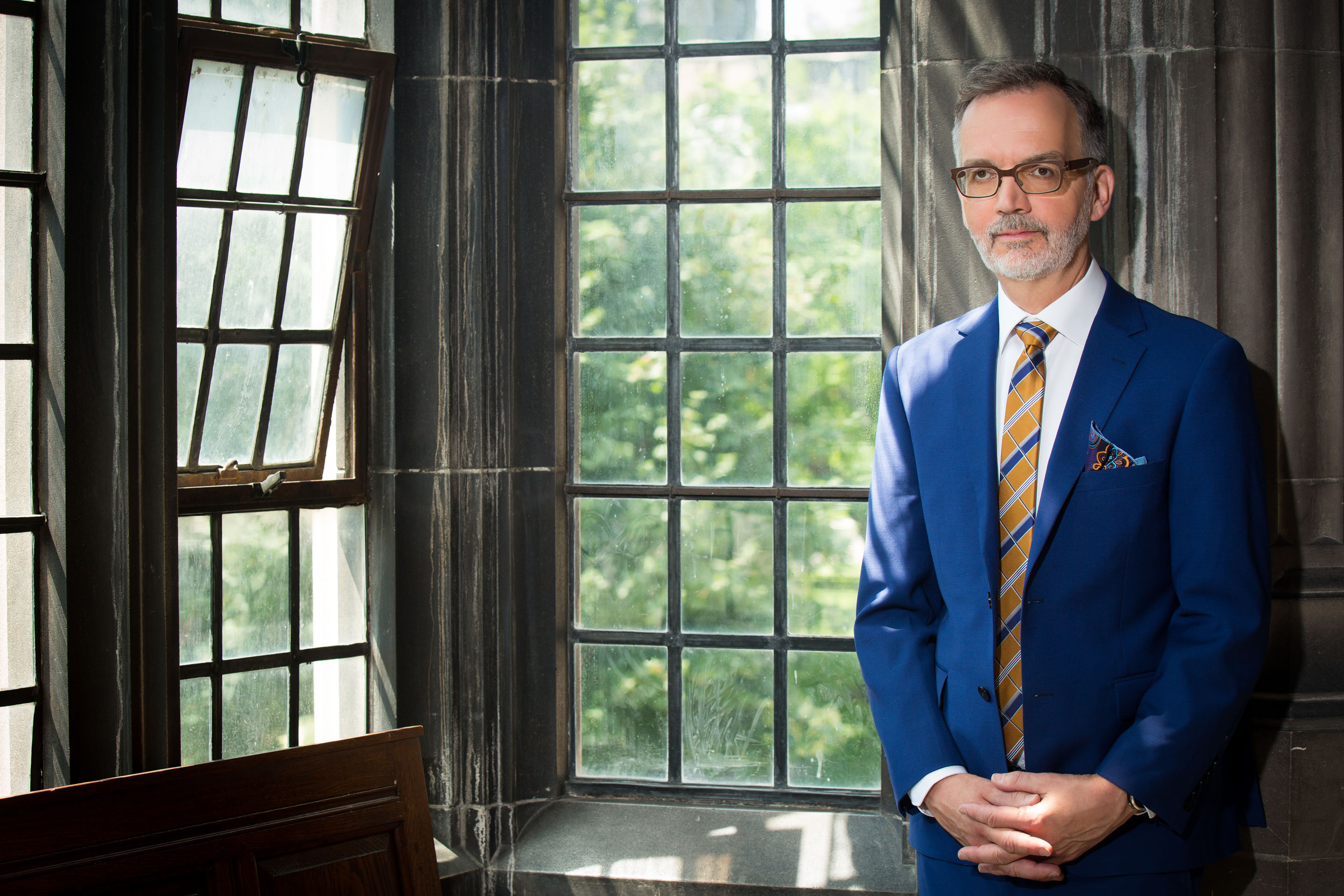
U of T Endocrinologist to Lead ‘Powerhouse’ Diabetes Research Network
A noted diabetes researcher and public health advocate from the Temerty Faculty of Medicine and the Dalla Lana School of Public Health has been selected lead a “powerhouse” research network that will impact the global fight against diabetes and other serious chronic diseases.
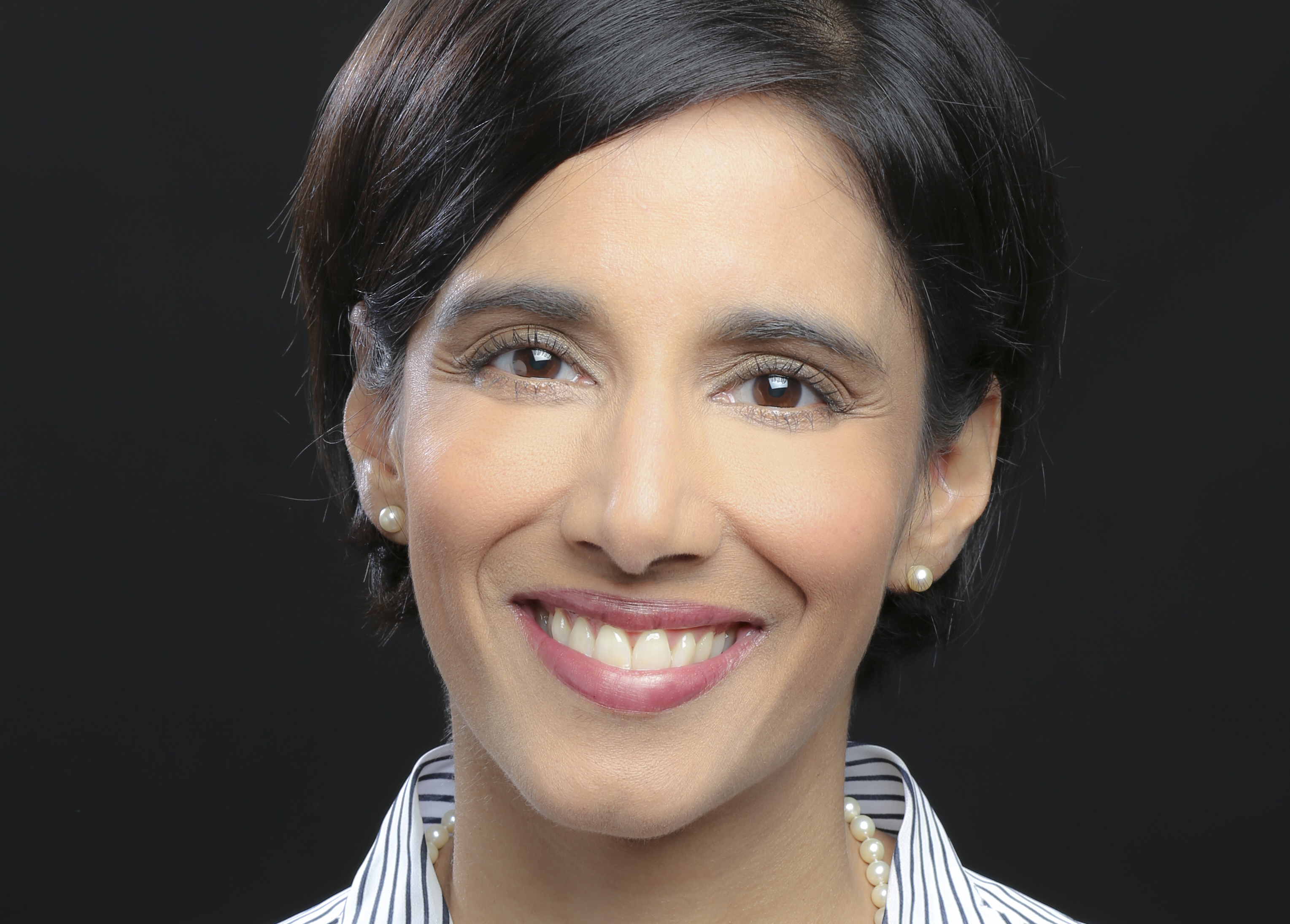
Physician and Associate Professor Lorraine Lipscombe of the Department of Medicine has been appointed director of the Novo Nordisk Network for Healthy Populations.
Lipscombe is a respected endocrinologist who holds appointments at Women’s College Hospital, where she is director of the hospital’s endocrinology division and is a senior scientist in the Women’s College Research Institute. Her research focuses on the prevention and improvement of care and outcomes for patients with diabetes, particularly women.
In February 2021, Novo Nordisk and the University of Toronto announced a $40-million investment to establish the Novo Nordisk Network for Healthy Populations. Based at U of T Mississauga, the network is a partnership between the Temerty Faculty of Medicine, the Dalla Lana School of Public Health and University of Toronto Mississauga. The network will focus on interdisciplinary collaboration to accelerate on-the-ground diabetes research, education and outreach.
“I am truly excited and honoured for the chance to lead this visionary network in demonstrating innovative and effective ways to make populations healthier,” Lipscombe said.
“The Network will afford an unprecedented opportunity to integrate and align expertise across multiple areas to establish a world-leading research program, through strategic collaborations between three U of T academic powerhouses and key community stakeholders in the City of Mississauga.”
“Being one of the most diverse cities in the world, with a mix of urban and suburban areas, and much higher rates of obesity and diabetes than the national average, Mississauga provides a unique environment to explore interventions that can be applied to a wide range of contexts around the world.”
The Network executive team welcomed news of Lipscombe’s appointment.
U of T Mississauga Vice-President and Principal Alexandra Gillespie lauded the incoming director as “a world-class researcher, proven team builder and dynamic leader known for sharing her expertise to empower the success and wellbeing of others.”
“With her guiding hand, the Network will achieve its goal of identifying and implementing strategies to prevent diabetes and diabetes complications in high risk and marginalized communities,” said Gillian Hawker, Sir John and Lady Eaton Professor and Chair of the Department of Medicine.
Lipscombe’s expertise in both clinical care and population health makes her “a superb choice to lead the network," said Professor Adalsteinn (Steini) Brown, Dean of the Dalla Lana School of Public Health, where Lipscombe teaches in the Institute for Health Policy, Management and Evaluation.
"As a clinician and an epidemiologist, she is particularly skilled at seeing the gaps between research and health delivery, and has developed an impressive track record in addressing them."
Gillespie also noted Lipscombe’s strategic planning skills.
“Professor Lipscombe will enable the Novo Nordisk Network to achieve its ambitious goals: to unite university divisions, hospitals, and community partners in the fight against diabetes and other serious chronic illnesses,” Gillespie said.
“Her leadership will benefit the health of so many people in Mississauga, Toronto, and around the world.”
The announcement of Lipscombe’s appointment comes at a momentous time for diabetes research, as U of T continues a year of celebrations marking the 100th anniversary of the discovery of insulin.
In June, the City of Mississauga announced that it will become the first Canadian municipality to join the Cities Changing Diabetes program. The Novo Nordisk-supported initiative will provide the City with tools, resources and partners – including the Network research hub – to prevent the rise of Type 2 diabetes in Mississauga.
About 420 million people worldwide live with diabetes. The World Health Organization estimates that the disease is responsible for more than 1.6 million deaths annually. Complications can lead to devastating health outcomes, such as blindness or limb amputation.
“The discovery of insulin is a monumental achievement for Canada and a beautiful example of the translation of research findings into life-altering benefits for people,” Lipscombe said.
“Despite this discovery and all the progress that has been made since then, diabetes remains a major burden on individuals, families, communities and health care systems around the world.”
“Much research has been done to recognize root causes of diabetes and its consequences and to identify effective interventions,” she continued.
“We must now act on this evidence, by shifting our focus from describing what might work to showing what does work.”

Optimize this page for search engines by customizing the Meta Title and Meta Description fields.
Use the Google Search Result Preview Tool to test different content ideas.
Select a Meta Image to tell a social media platform what image to use when sharing.
If blank, different social platforms like LinkedIn will randomly select an image on the page to appear on shared posts.
Posts with images generally perform better on social media so it is worth selecting an engaging image.
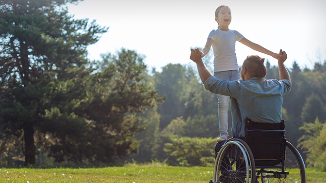
Balancing Act
Researchers have created a unique rehabilitation program that can help some of the 86,000 Canadians who live with a spinal cord injury achieve greater independence.
Spinal cord injuries can be ‘complete’—leading to the total loss of movement control at and below the site of injury—or ‘incomplete’—when some ability to move is retained.
Individuals who live with an incomplete spinal cord injury can regain some standing and walking abilities; however, they often have trouble balancing, which makes it difficult for them to stand without support and puts them at a high risk of falling.
To address this issue, a team led by Kristin Musselman, a professor of physical therapy, and Kei Masani, a professor of clinical engineering in the Institute of Biomedical Engineering, explored whether a new balance training program could improve perceived standing ability and confidence in individuals with an incomplete spinal cord injury. Musselman and Masani are also researchers at the KITE Research Institute.
This study builds on previous research in which the team evaluated the innovative four-week balance training program. The program involves balance exercises combined with performance feedback, which is displayed on a computer monitor. During the exercises, electrical stimulation is applied to the ankle muscles to induce contractions that help the participants maintain balance. Repeated exercise with muscle stimulation helps to retrain the nervous system to achieve greater balance control.
“Our initial study showed that this program improves balance, but it did not give us any insights into whether the participants felt that the program was beneficial,” explains Musselman. “It is important to consider the perceived benefits of a training program to ensure that the programs we are developing are meaningful to patients. Patients are more likely to participate in rehabilitation if they find it valuable.”
To explore participants’ perspectives, the researchers interviewed five individuals who completed the training program. The participants answered questions about their goals and expectations, and how the program impacted their life, balance confidence and risk of falling.
Participants reported that the program increased their range of motion and muscle strength and decreased muscle spasms and pain. They also reported feeling more independent and better able to complete daily tasks such as cooking, self-dressing and using the washroom.
 Importantly, participants gained confidence in their physical abilities and became more willing to try new activities. They also felt that they could continue to improve and were motivated to carry on with balance training.
“We knew that our balance training program benefits patients physically, but now we see that it also benefits them psychologically,” says Musselman. “This program can help individuals with an incomplete spinal cord injury gain the confidence needed to be more independent and carry out meaningful daily activities.”
Although this rehabilitation program is not yet available in clinics, the researchers have recently received funding to develop a new version of it that is appropriate for clinical practice. They hope to test this new program in patients with spinal cord injury, as well as stroke, in the next few years.
This work was supported by the University of Toronto EMHSeed Program and the UHN Foundation.
Source: University Health Network
Researchers have created a unique rehabilitation program that can help some of the 86,000 Canadians who live with a spinal cord injury achieve greater independence.
Spinal cord injuries can be ‘complete’—leading to the total loss of movement control at and below the site of injury—or ‘incomplete’—when some ability to move is retained.
Individuals who live with an incomplete spinal cord injury can regain some standing and walking abilities; however, they often have trouble balancing, which makes it difficult for them to stand without support and puts them at a high risk of falling.
To address this issue, a team led by Kristin Musselman, a professor of physical therapy, and Kei Masani, a professor of clinical engineering in the Institute of Biomedical Engineering, explored whether a new balance training program could improve perceived standing ability and confidence in individuals with an incomplete spinal cord injury. Musselman and Masani are also researchers at the KITE Research Institute.
This study builds on previous research in which the team evaluated the innovative four-week balance training program. The program involves balance exercises combined with performance feedback, which is displayed on a computer monitor. During the exercises, electrical stimulation is applied to the ankle muscles to induce contractions that help the participants maintain balance. Repeated exercise with muscle stimulation helps to retrain the nervous system to achieve greater balance control.
“Our initial study showed that this program improves balance, but it did not give us any insights into whether the participants felt that the program was beneficial,” explains Musselman. “It is important to consider the perceived benefits of a training program to ensure that the programs we are developing are meaningful to patients. Patients are more likely to participate in rehabilitation if they find it valuable.”
To explore participants’ perspectives, the researchers interviewed five individuals who completed the training program. The participants answered questions about their goals and expectations, and how the program impacted their life, balance confidence and risk of falling.
Participants reported that the program increased their range of motion and muscle strength and decreased muscle spasms and pain. They also reported feeling more independent and better able to complete daily tasks such as cooking, self-dressing and using the washroom.
Importantly, participants gained confidence in their physical abilities and became more willing to try new activities. They also felt that they could continue to improve and were motivated to carry on with balance training.
“We knew that our balance training program benefits patients physically, but now we see that it also benefits them psychologically,” says Musselman. “This program can help individuals with an incomplete spinal cord injury gain the confidence needed to be more independent and carry out meaningful daily activities.”
Although this rehabilitation program is not yet available in clinics, the researchers have recently received funding to develop a new version of it that is appropriate for clinical practice. They hope to test this new program in patients with spinal cord injury, as well as stroke, in the next few years.
This work was supported by the University of Toronto EMHSeed Program and the UHN Foundation.
Source: University Health Network
Optimize this page for search engines by customizing the Meta Title and Meta Description fields.
Use the Google Search Result Preview Tool to test different content ideas.
Select a Meta Image to tell a social media platform what image to use when sharing.
If blank, different social platforms like LinkedIn will randomly select an image on the page to appear on shared posts.
Posts with images generally perform better on social media so it is worth selecting an engaging image.
Congratulations to the Class of 2021
Yesterday, I had the opportunity to congratulate the MD Class of 2021, which officially received their degrees. They will soon be followed by graduates of other programs from the Temerty Faculty of Medicine, including students from Medical Radiation Sciences and research-based graduate programs, who will join the rest of the University of Toronto in marking Spring Convocation on June 23. Due to the COVID-19 pandemic, these students have experienced a learning environment that few could have envisioned. It has meant lessons shared from a distance, a clinical learning environment under extreme pressure, and thesis defences conducted online instead of in-person. One skill many of these learners have certainly now mastered: donning and doffing personal protective equipment.
Our newest graduates begin the next stages of their lives at a time of uncertainty and change. COVID-19 has presented challenges to our society, to our economy, but most importantly, to how we deliver healthcare in a safe, equitable, and meaningful way. Our communities and society are also wrestling with longstanding injustices and an imperative to meaningfully address historical inequities. As healthcare professionals, we have a role to play in this vital work. Such uncertainty and change can cause anxiety, but they can also present tremendous opportunities.
Just over one hundred years ago, the world was recovering from the Spanish Flu pandemic and World War One. Societies, international alliances, economic order, and global health all needed to be rebuilt. There was ambiguity and, no doubt, a great sense of weariness. Yet, new possibilities were being realized. For example, in a lab at the University of Toronto, insulin was about to be discovered. Significant challenges sometimes present us with great opportunities to reimagine our world for the better, and our quest for discovery continues regardless of crises that may surround us. Herein lies the challenge before the Class of 2021, and I am confident that they can meet the demands of this moment
I hope that the Class of 2021 feels that we – the faculty and staff of Temerty Medicine – have been there to support them through these trying times. Especially now throughout the pandemic, I have seen our faculty and staff consistently go above and beyond to provide a sympathetic ear, helpful suggestions, or alternatives that have made the journey our learners are on a little easier. And while they may graduate, they will always remain members of Temerty Medicine. They join a network of over 60,000 healthcare leaders who are valued members of our community and reinforce the Temerty Faculty of Medicine’s world-class reputation. I hope they return to our campuses often as they pursue a lifelong commitment to learning.
On behalf of the Temerty Faculty of Medicine, I want to extend my heartfelt congratulations to the Class of 2021. And I want to thank all of the people who have helped our graduates reach this milestone; the family and friends, the classmates and alumni, and the faculty and staff. No graduate reaches this moment by themselves, which is why it’s essential that we – as a community – celebrate this collective achievement.
Trevor Young
Dean, Temerty Faculty of Medicine
Vice Provost, Relations with Health Care Institutions
University of Toronto
Optimize this page for search engines by customizing the Meta Title and Meta Description fields.
Use the Google Search Result Preview Tool to test different content ideas.
Select a Meta Image to tell a social media platform what image to use when sharing.
If blank, different social platforms like LinkedIn will randomly select an image on the page to appear on shared posts.
Posts with images generally perform better on social media so it is worth selecting an engaging image.
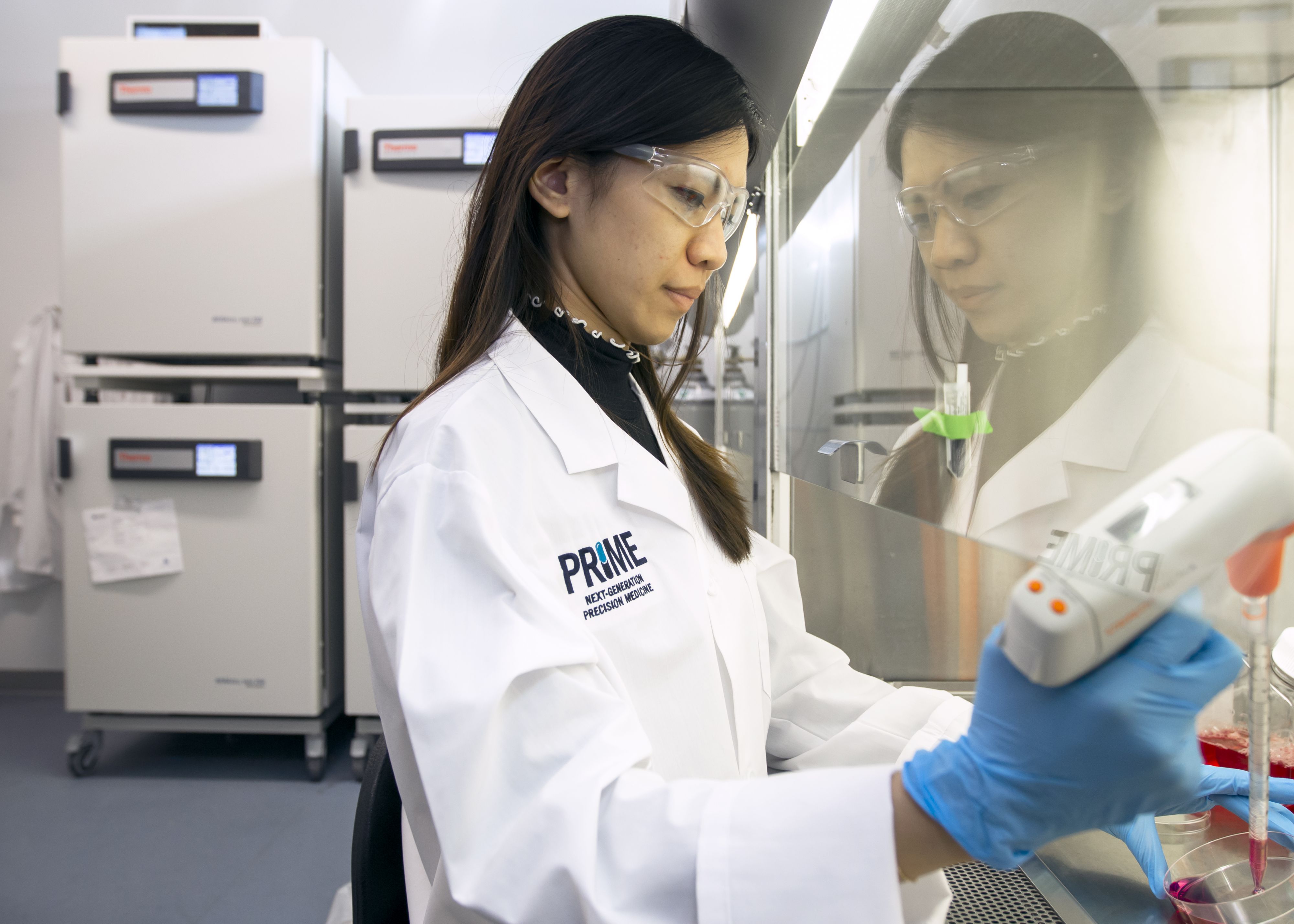
U of T’s New Precision Medicine Program Offers Training in Cross-Disciplinary Science
A new graduate education program at the University of Toronto will provide PhD students with advanced education in interdisciplinary biomedical research.
The Collaborative Specialization in Next-Generation Precision Medicine opened for enrolment this week and will offer courses this fall at the Temerty Faculty of Medicine, the Leslie Dan Faculty of Pharmacy, the Faculty of Arts & Science and the Faculty of Applied Science and Engineering.
The program’s courses and modules are linked to PRiME, a cross-disciplinary research effort launched by the University in 2019 as an Institutional Strategic Initiative (ISI) with a focus on precision medicine that spans physical and life sciences, and engineering.
“Over the last couple of years, the value of PRiME’s research activities has become apparent and new collaborations are blossoming,” said Rob Batey, professor and chair of the department of chemistry in Arts & Science and a researcher with PRiME. “It’s only natural that PRiME would turn its attention to education and introducing cross-disciplinary research as early as possible in a graduate student’s career.”
The new program is run through PRiME and is the first collaborative specialization based on an ISI at the University. There are more than 40 such specializations at U of T, which provide a multidisciplinary experience while allowing students to complete a related degree.
PRiME is led by Professor Shana Kelley at the Leslie Dan Faculty of Pharmacy and brings together 75 investigators and their trainees to advance research in disease biology, diagnostics and drug discovery. The new collaborative specialization draws 16 core faculty members from that group including Christine Allen, a professor at the Leslie Dan Faculty of Pharmacy who is U of T’s associate vice-president and vice-provost of strategic initiatives.
“There is a need for innovative and interdisciplinary educational offerings in this area,” said Allen. “These will serve to solidify the foundational concepts that underpin precision medicine and drug development and benefit our trainees once they transition to the workforce.”
Allen said the specialization is a sign that PRiME is succeeding as a research initiative.
“We always expected that once we created the research networks the educational offerings would follow. We brought people together through supporting the research and now they want to teach together,” said Allen. “It’s an indication that PRiME is succeeding and growing, and people want to be part of it.”
Research and education in next-generation, precision medicine requires expertise in diverse areas including biologics, omics, molecular chemistry, liquid biopsy, nanomedicine and biology-on-a-chip. Work in the field is by necessity collaborative and interdisciplinary.
Geordi Frere is a second-year PhD student in chemistry at U of T Mississauga, co-supervised by Professors Scott Prosser and Patrick Gunning. He said that interdisciplinary courses are appealing, especially for the access they offer to a large network of researchers with various expertise.
“It’s a really great chance to expand the avenues of research that you can explore. There’s an amazing wealth of expertise in different faculties at U of T that PRiME helps you tap into,” Frere said. “The collaborative specialization will help students familiarize themselves with these resources and learn how to access them efficiently.”
Students who are interested in applying to the Collaborative Specialization in Next-Generation Precision Medicine can visit education.prime.utoronto.ca for more information.
Optimize this page for search engines by customizing the Meta Title and Meta Description fields.
Use the Google Search Result Preview Tool to test different content ideas.
Select a Meta Image to tell a social media platform what image to use when sharing.
If blank, different social platforms like LinkedIn will randomly select an image on the page to appear on shared posts.
Posts with images generally perform better on social media so it is worth selecting an engaging image.
Job Shadowing Goes Virtual for Mississauga MD Program

When you can’t go to a job shadowing opportunity at the hospital, can the opportunity come to you?
That’s the question posed by one enterprising learner who created a unique initiative for MD students at Temerty Medicine.
Max Solish is a second-year student in the Mississauga Academy of Medicine (MAM). Solish and his classmates planned to observe different medical specialties through shadowing, but COVID-19 safety protocols kept early-year students on the sidelines.
Undaunted, Solish reached out to Trillium Health Partners (THP) with a novel solution – a virtual shadowing opportunity that would grant MAM learners the chance to watch live surgical procedures by videoconference.
In the third year of the program, MD students begin formal clinical rotations to gain experience with different medical specialties. The process, known as clerkship, helps students determine which fields they want to specialize in after they complete their medical degrees.
But it’s never too early to start the process, which is why Solish and his second-year classmates were anxious to seize any opportunity to learn more about the various medical specialties open to them.
Solish connected with Bryan Abankwah, THP’s manager of education, and Michael Weinberg, (MD ’90, PGME ’95), an assistant professor of plastic and reconstructive surgery and physician education lead in THP’s plastic surgery division. Abankwah and Weinberg were enthusiastic about the idea.
“Exposure to specialties in a pre-med environment is important,” says Weinberg. “Learners must make a crucial decision about specialization very early, and that can be tough when the pandemic prohibits them from being there in person.”
The virtual shadowing initiative is a first for both THP and MAM.
With a secure videoconference platform and careful attention to patient privacy protocols, the team was ready to pilot the first-ever virtual visit.
In early May, six second-year MAM students tuned in to watch Weinberg perform a series of on-camera procedures, including hand surgery, mole removal and a skin cancer lesion excision. The outpatient surgeries, which Weinberg deems the “bread-and-butter” of his specialty, are performed relatively quickly and under local anaesthetic. Two-way communication throughout ensured student learners could ask questions throughout the process and join Weinberg for an informal chat afterwards.
“It was an opportunity to see what it’s all about, and get a feel for the work,” says Weinberg. “I hope I demystified it and we gave the learners some exposure in a collaborative environment they couldn’t have gotten otherwise.”
Based on the success of the pilot project, a second session took place with a new group of learners on June 1. Solish hopes to schedule more virtual opportunities over the coming months so all interested MAM learners can participate.
According to Abankwah, there is growing enthusiasm for the project.
“We’ve had positive inquiries from other faculty members who want to participate,” he says. “We’re exploring the possibility of expanding the scope as we look at future virtual learning opportunities for students.”
While the demonstrations are open only to MAM learners affiliated with THP, Solish hopes other hospital partners will take notice and offer similar opportunities to students across Temerty Medicine’s four academies.
“Ultimately, you need more than a two-hour video experience, but virtual shadowing is a gateway to learn more about that specialty,” Solish says.
For his part, Weinberg looks forward to welcoming the next group of virtual observers.
“In unpredictable times, when we’re challenged on every level, we can be resourceful and find innovative and fun new ways to teach.”
Optimize this page for search engines by customizing the Meta Title and Meta Description fields.
Use the Google Search Result Preview Tool to test different content ideas.
Select a Meta Image to tell a social media platform what image to use when sharing.
If blank, different social platforms like LinkedIn will randomly select an image on the page to appear on shared posts.
Posts with images generally perform better on social media so it is worth selecting an engaging image.
Blake Eligh
Celebrating 75 Years of Scientific and Medical Art
Temerty Medicine’s Master of Science in Biomedical Communications (MScBMC) program marks a milestone this June with disruptors, an exhibition of medical and scientific art spanning the program’s 75 years.
The online retrospective is part of a month-long schedule of BMC 75 events that wrap the program’s anniversary year.
And there is much to celebrate.
The MScBMC program is the sole Canadian program — and one of just four in North America —to offer graduate level training in visual science communications. Founded in 1945 as a certificate program called Art as Applied to Medicine (AAM), it became a bachelor of science degree (BScAAM) in the 1960s before evolving yet again into a two-year graduate studies program (MScBMC) in the 1990s when it was renamed biomedical communications.
Now based at U of T’s Mississauga campus, the program provides highly specialized training to graduate students to create illustrations and visualizations to promote understanding, communication and advancement of science and medicine.
Used to communicate to a wide variety of audiences, inlcuding scientists, medical health professionals and lay audiences, the work is published in journals and textbooks, used in teaching and marketing materials, and even appears in courtrooms to illustrate testimony by expert medical witnesses.
EVOLUTION OF SCIENCE COMMUNICATION

“The focus of our work and the tools at our disposal have evolved tremendously over the past 75 years but the ethos of our practice remains the same,” says program director and alumna Jodie Jenkinson (MScBMC 1998).
“At its core, biomedical communications is a problem-solving activity involving the deconstructing of structures and processes and the identification of visual communication strategies to convey these phenomena with clarity and accuracy.”
Professor emerita Margot Mackay agrees. Mackay (BScAAM 1968) joined the program as a student in 1964 and launched a career creating step-by-step illustrations to help surgeons communicate their work to other practitioners as well as to patients and their families.
Mackay and her contemporaries worked in carbon dust, watercolour or pen and ink for journal publications, while teaching relied on projection media like 35 mm slides and video production.
“In the early days, we didn’t have computers, so you had to fine-tune your drawing skills,” she says.
Now retired, Mackay continues to critique student work. She notes that contemporary illustrators also train to create visualizations with digital animation platforms, tablet technology and augmented reality.
While tools of the trade have evolved, so too has the subject matter. Advancements in medical technology and scientific knowledge ensure there’s always new and different information to communicate.
Mackay recalls perching atop a special step ladder with a bird’s eye view of the operating room to make drawings of some of the earliest lung transplant procedures performed in Toronto.
“The content is now at the cellular or micro level, showing the reactions of proteins and parts of the cells,” Mackay says.
“That’s where the research is. Illustrators have always been part of the front line to assist scientists in communicating that research.”
THE ART AND SCIENCE OF DISRUPTION

“If the past year has shown us anything, it’s the importance of scientific communication as a vehicle for informing and engaging the public,” says Jenkinson.
“The pandemic derailed initial plans to hold a gala event but gave us time to reflect on the profession and our role in society.”
The anniversary exhibition, titled disruptors, features contemporary and archival work from students, faculty and alumni that explores the impact of disruption on the content of science communication and the techniques used by the communicators.
Curated by first-year MScBMC students Mimi (Yuejun) Guo and Shay Saharan, disruptors is hosted on a digital platform that mimics a physical gallery space. Works are displayed on virtual walls and include clickable pop-ups with information about the artist and their work.
The exhibit is divided into three sections that explore how societal changes, advances in science and medicine and how new visualization tools have influenced the field. A new phase will be released each Monday throughout the month, beginning May 31. The full catalogue will be on display during the week of June 21 to 27.

Visitors to the gallery will find archival images such as carbon dust illustrations from the ground-breaking Grant’s Atlas of Anatomy and images by program founder Maria Wishart, who documented the response to the smallpox vaccination in the 1930s. Their work is displayed alongside contemporary digital images and videos created by recent alumni and current students.
Guo says it was exciting to curate the retrospective. “We can learn from each other and admire each other’s work,” she says.
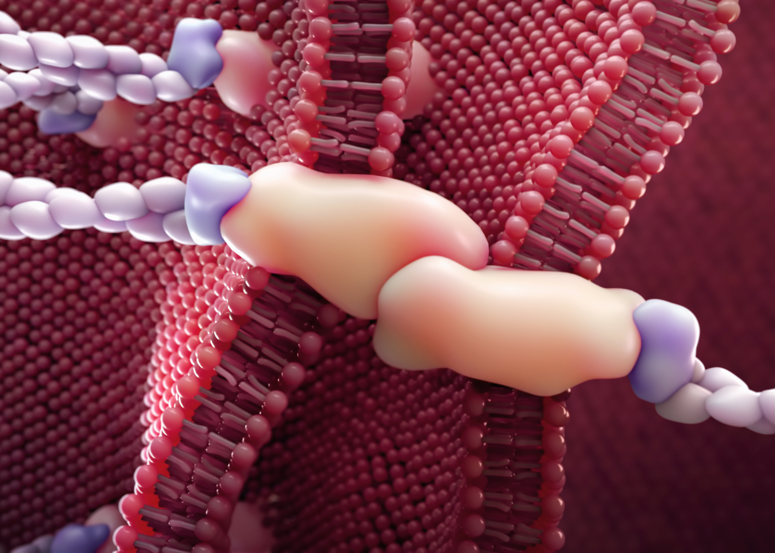
Alumnus Derek Ng (MScBMC 2015, now an assistant professor with the program who teaches molecular visualization and interactive media design, says his favourite piece in the exhibition is an animation of the SARS-CoV-2 virus particle.
The animation was created by another alumnus, Nick Woolridge (BScBMC 1991), who is now an associate professor and past director of the program. It has been used internationally by public health leaders to communicate the science of COVID-19.
“It shows the virus and cuts away to show the internal architecture,” Ng says. “It’s a great visualization of molecular science.”
The exhibit also includes an online scavenger hunt to encourage participats to explore the exhibit, with a chance to win copies of the commemorative catalogue and limited-edition, double-sided jigsaw puzzle.
The catalogue and the puzzle feature the 1943 illustration Dissection of the Facial Nerve by one of the program’s first graduates, Marguerite Drummond (AAM 1946) and Blood-Brain Barrier by alumna and biomedical artist Hang Yu Lin (MScBMC 2020), are also available for purchase.
“It’s cool to see how far the profession has come over 75 years,” says Saharan. “You can see the changes in technique and technology, as well as what we’re capable of doing right now and how that’s changing the skills we need to learn to make science accessible.
disruptors runs online until June 27. Visit the MScBMC News and Events page for more information about the exhibit, alumni workshops and other BMC 75 events.
Temerty Medicine’s Master of Science in Biomedical Communications (MScBMC) program marks a milestone this June with disruptors, an exhibition of medical and scientific art spanning the program’s 75 years.
The online retrospective is part of a month-long schedule of BMC 75 events that wrap the program’s anniversary year.
And there is much to celebrate.
The MScBMC program is the sole Canadian program — and one of just four in North America —to offer graduate level training in visual science communications. Founded in 1945 as a certificate program called Art as Applied to Medicine (AAM), it became a bachelor of science degree (BScAAM) in the 1960s before evolving yet again into a two-year graduate studies program (MScBMC) in the 1990s when it was renamed biomedical communications.
Now based at U of T’s Mississauga campus, the program provides highly specialized training to graduate students to create illustrations and visualizations to promote understanding, communication and advancement of science and medicine.
Used to communicate to a wide variety of audiences, inlcuding scientists, medical health professionals and lay audiences, the work is published in journals and textbooks, used in teaching and marketing materials, and even appears in courtrooms to illustrate testimony by expert medical witnesses.
EVOLUTION OF SCIENCE COMMUNICATION
“The focus of our work and the tools at our disposal have evolved tremendously over the past 75 years but the ethos of our practice remains the same,” says program director and alumna Jodie Jenkinson (MScBMC 1998).
“At its core, biomedical communications is a problem-solving activity involving the deconstructing of structures and processes and the identification of visual communication strategies to convey these phenomena with clarity and accuracy.”
Professor emerita Margot Mackay agrees. Mackay (BScAAM 1968) joined the program as a student in 1964 and launched a career creating step-by-step illustrations to help surgeons communicate their work to other practitioners as well as to patients and their families.
Mackay and her contemporaries worked in carbon dust, watercolour or pen and ink for journal publications, while teaching relied on projection media like 35 mm slides and video production.
“In the early days, we didn’t have computers, so you had to fine-tune your drawing skills,” she says.
Now retired, Mackay continues to critique student work. She notes that contemporary illustrators also train to create visualizations with digital animation platforms, tablet technology and augmented reality.
While tools of the trade have evolved, so too has the subject matter. Advancements in medical technology and scientific knowledge ensure there’s always new and different information to communicate.
Mackay recalls perching atop a special step ladder with a bird’s eye view of the operating room to make drawings of some of the earliest lung transplant procedures performed in Toronto.
“The content is now at the cellular or micro level, showing the reactions of proteins and parts of the cells,” Mackay says.
“That’s where the research is. Illustrators have always been part of the front line to assist scientists in communicating that research.”
THE ART AND SCIENCE OF DISRUPTION
“If the past year has shown us anything, it’s the importance of scientific communication as a vehicle for informing and engaging the public,” says Jenkinson.
“The pandemic derailed initial plans to hold a gala event but gave us time to reflect on the profession and our role in society.”
The anniversary exhibition, titled disruptors, features contemporary and archival work from students, faculty and alumni that explores the impact of disruption on the content of science communication and the techniques used by the communicators.
Curated by first-year MScBMC students Mimi (Yuejun) Guo and Shay Saharan, disruptors is hosted on a digital platform that mimics a physical gallery space. Works are displayed on virtual walls and include clickable pop-ups with information about the artist and their work.
The exhibit is divided into three sections that explore how societal changes, advances in science and medicine and how new visualization tools have influenced the field. A new phase will be released each Monday throughout the month, beginning May 31. The full catalogue will be on display during the week of June 21 to 27.
Visitors to the gallery will find archival images such as carbon dust illustrations from the ground-breaking Grant’s Atlas of Anatomy and images by program founder Maria Wishart, who documented the response to the smallpox vaccination in the 1930s. Their work is displayed alongside contemporary digital images and videos created by recent alumni and current students.
Guo says it was exciting to curate the retrospective. “We can learn from each other and admire each other’s work,” she says.
Alumnus Derek Ng (MScBMC 2015, now an assistant professor with the program who teaches molecular visualization and interactive media design, says his favourite piece in the exhibition is an animation of the SARS-CoV-2 virus particle.
The animation was created by another alumnus, Nick Woolridge (BScBMC 1991), who is now an associate professor and past director of the program. It has been used internationally by public health leaders to communicate the science of COVID-19.
“It shows the virus and cuts away to show the internal architecture,” Ng says. “It’s a great visualization of molecular science.”
The exhibit also includes an online scavenger hunt to encourage participats to explore the exhibit, with a chance to win copies of the commemorative catalogue and limited-edition, double-sided jigsaw puzzle.
The catalogue and the puzzle feature the 1943 illustration Dissection of the Facial Nerve by one of the program’s first graduates, Marguerite Drummond (AAM 1946) and Blood-Brain Barrier by alumna and biomedical artist Hang Yu Lin (MScBMC 2020), are also available for purchase.
“It’s cool to see how far the profession has come over 75 years,” says Saharan. “You can see the changes in technique and technology, as well as what we’re capable of doing right now and how that’s changing the skills we need to learn to make science accessible.
disruptors runs online until June 27. Visit the MScBMC News and Events page for more information about the exhibit, alumni workshops and other BMC 75 events.
Optimize this page for search engines by customizing the Meta Title and Meta Description fields.
Use the Google Search Result Preview Tool to test different content ideas.
Select a Meta Image to tell a social media platform what image to use when sharing.
If blank, different social platforms like LinkedIn will randomly select an image on the page to appear on shared posts.
Posts with images generally perform better on social media so it is worth selecting an engaging image.
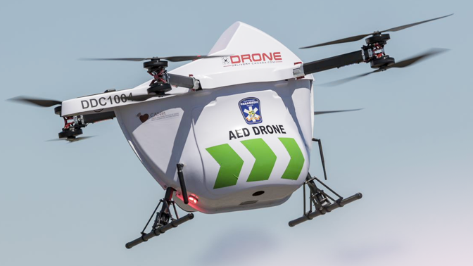
Help from Above
The use of drones to deliver of goods and services is getting closer to reality every day. In health care, drones could be used to rapidly deliver life-saving automatic external defibrillators (AED) to people experiencing cardiac arrest.
Timothy Chan, a professor of mechanical and industrial engineering and member of the Temerty Centre for AI Research and Education in Medicine (T-CAIREM) used historical Ontario data, which showed when and where cardiac arrests had occurred, to reveal that there are substantial benefits to using drones to deliver AEDs.
AEDs are available at many public venues, such as subway stations, stadiums and universities, but are less likely to be available close to someone suffering cardiac arrest in residential and rural settings. Drones could deliver an AED in these cases; however, there are trade-offs. It would be a waste of resources to send a drone in response to a 911 call if an ambulance arrived sooner.
To address these issues, a research team led by Chan developed a machine learning-based system to decide when to optimally dispatch a drone. The team tested different decision-making approaches using a simulation based on data from Peel region between 2015 and 2019.
A simple rule — to always send a drone — would have improved response times by approximately two to three minutes, suggesting that there is strong potential for improving patient outcomes in residential settings like Peel. Each minute gained could improve the odds of survival by 10% based on a common rule of thumb.
The machine learning optimized rules were able to achieve nearly identical improvements in response time, while dispatching 30% fewer drones. The advanced decision-making approach could help municipalities implement drone-delivered AEDs to save lives, at a reduced cost compared to sending them in response to every call.
“The high accuracy of our dispatch rules is bolstered by the fact that even when an incorrect decision was made, the time difference it would have taken an ambulance to arrive was relatively small,” Chan, who is also an affiliate scientist at the Techna Institute for the Advancement of Technology for Health , says of the results. “For a drone-delivered AED initiative to succeed, other barriers must be overcome. The concept requires community buy-in: not just the acceptance of drones flying through the area, but the acceptance of delivered AEDs. This would require targeted education and social marketing campaigns. The logistics of operating and maintaining a drone network must also be developed,” he concludes.
This work was supported by the Cardiac Arrhythmia Network of Canada and Zoll Medical Corporation. TY Chan holds a Tier 2 Canada Research Chair in Novel Optimization and Analytics in Health. A conflict-of-interest was declared in this study for Dr. Cheskes, who received funding from Zoll Medical and sits on the Advisory Board of Drone Delivery Canada.
Optimize this page for search engines by customizing the Meta Title and Meta Description fields.
Use the Google Search Result Preview Tool to test different content ideas.
Select a Meta Image to tell a social media platform what image to use when sharing.
If blank, different social platforms like LinkedIn will randomly select an image on the page to appear on shared posts.
Posts with images generally perform better on social media so it is worth selecting an engaging image.
Honouring Those Lost and Acting for Change
At the Temerty Faculty of Medicine we support President Gertler’s statement regarding the discovery of remains of 215 Indigenous children found at a former residential school in Tk’emlúps te Secwépemc territory. We cannot imagine the suffering and pain those children endured and the trauma their families and communities continue to face.
We are aware that this discovery has occurred in the context of the inquest into the death of Joyce Echaquan and the racist treatment she received by healthcare professionals prior to her death.
Tragically, while neither of these events is unprecedented for Indigenous peoples, both contribute to the cumulative trauma that generations of Indigenous peoples have suffered since the arrival of settlers on Turtle Island. These events are reminders of how much further we have to go to address the horrifying and ongoing impacts of settler colonialism on the rights, experiences, and health and well-being of Indigenous peoples.
We recognize the individual and collective grief Indigenous communities are experiencing right now. At this time, we would like to encourage our non-Indigenous community members to express solidarity with and compassion for Indigenous community members, and to commit to advancing truth and reconciliation in all our institutions.
At the Temerty Faculty, in accordance with the Calls to Action of the Truth and Reconciliation Commission of Canada, our obligation and commitment are to work with and seek ongoing guidance from Indigenous communities to ensure a present and a future – in our education, research, and advocacy – where these events are neither forgotten nor repeated in our healthcare and academic environments.
To support this commitment, the Faculty is actively working to improve the Indigenous learner experience; strengthen admissions pathways; deepen the Indigenous health curriculum; advance Indigenous faculty and staff; offer Indigenous governance and guidance for leadership; strengthen Indigenous community partnerships; and educate around cultural safety and anti-racist practice.
For Indigenous members of the Temerty Faculty community seeking connection and support, there are resources available:
- Please contact indigenoushealth.support@utoronto.ca or First Nations House at U of T.
- An Indian Residential Schools Crisis Line (1-866-925-4419) is available 24 hours a day for anyone experiencing pain or distress because of his or her residential school experience.
- Native Women’s Association of Canada offers in-house elder support at 888-664-7808.
- The Government of Canada has a Hope for Wellness line at 1-855-242-3310 or connect to the online chat at hopeforwellness.ca.
- Anishnawbe Health Toronto also offers support.
Together we must honour those lost and act collectively to bring about positive change, better health and greater well-being for all Indigenous peoples.
Optimize this page for search engines by customizing the Meta Title and Meta Description fields.
Use the Google Search Result Preview Tool to test different content ideas.
Select a Meta Image to tell a social media platform what image to use when sharing.
If blank, different social platforms like LinkedIn will randomly select an image on the page to appear on shared posts.
Posts with images generally perform better on social media so it is worth selecting an engaging image.
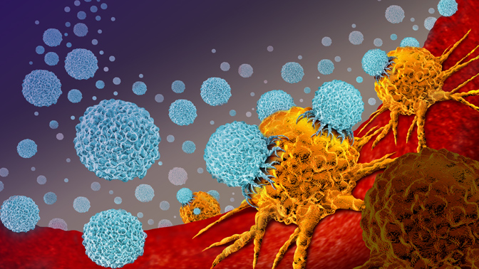
Staying On-Target
As a cancer becomes more aggressive, it can gain the ability to spread to other parts of the body. When this happens, it becomes harder to treat using targeted treatments, such as surgical removal and radiotherapy.
However, a recent study aimed to expand the opportunities for curing the disease by unveiling a new gray zone for prostate cancer patients — a state in which the cancer has begun to spread but can still be targeted.
Cells from the prostate have a specific molecule on their surfaces called Prostate Specific Membrane Antigen (PSMA). When cancer forms in the prostate, the resulting cancer cells also have PSMA on their surfaces. This molecule can be targeted using an imaging agent and visualized using positron emission tomography (PET). This approach can be used to find prostate cancer cells, even if they have spread to other parts of the body.
UHN has developed the ability to produce this imaging agent locally through CanProbe — a joint initiative between UHN and the Centre for Probe Development and Commercialization — which provides access to specialized expertise and facilities for the development of new radiopharmaceuticals.
A team led by Alejandro Berlin, a professor of radiation oncology and affiliated scientist with the Techna Institute was the first to apply this novel technology at UHN through a clinical trial involving 87 prostate cancer patients whose disease had returned. The conventional treatments aimed at curing these patients had been exhausted—and after prostate cancer returns and has spread, oncologists typically can no longer use targeted therapies. Instead, they resort to chemotherapy or hormone therapy — treatments that can control the disease for prolonged intervals but have little hope of eradicating the cancer.

The study aimed to use the advanced imaging approach to better identify the degree of prostate cancer spread and to see whether some of these patients could still benefit from targeted treatment.
Using the PSMA-PET method, the team found that 71% of patients had limited spread of cancer cells — which they termed “oligorecurrent disease” — versus just 22% with widespread disease. The trial then used the imaging results showing precisely where the cancer had spread so that radiation therapy or surgery could be used.
The targeted treatments were able to control the cancer in a majority of patients, and were able to completely remove the evidence of cancer in about one in five patients.
This study not only adds to the emerging evidence demonstrating the value of the PSMA imaging in the management prostate cancer, but it also expands potential use of curative treatments — particularly for those with a limited degree of spread (i.e., oligorecurrent cancer). The research team is now helping to advance larger international trials that will determine further individualized treatment strategies.
“This was a collaborative effort between the genitourinary site group at the Princess Margaret Cancer Centre and the Joint Department of Medical Imaging under the auspices and vision established by the Techna Institute. By enabling us to apply these cutting-edge approaches, this study has benefited and continues to benefit our patients,” concludes Berlin.
This work was supported by the Terry Fox Research Institute, Abbvie CARO Uro-Oncologic Radiation Awards, the Astellas Prostate Cancer Innovation Fund, the University of Toronto and The Princess Margaret Cancer Foundation.
Source: University Health Network
As a cancer becomes more aggressive, it can gain the ability to spread to other parts of the body. When this happens, it becomes harder to treat using targeted treatments, such as surgical removal and radiotherapy.
However, a recent study aimed to expand the opportunities for curing the disease by unveiling a new gray zone for prostate cancer patients — a state in which the cancer has begun to spread but can still be targeted.
Cells from the prostate have a specific molecule on their surfaces called Prostate Specific Membrane Antigen (PSMA). When cancer forms in the prostate, the resulting cancer cells also have PSMA on their surfaces. This molecule can be targeted using an imaging agent and visualized using positron emission tomography (PET). This approach can be used to find prostate cancer cells, even if they have spread to other parts of the body.
UHN has developed the ability to produce this imaging agent locally through CanProbe — a joint initiative between UHN and the Centre for Probe Development and Commercialization — which provides access to specialized expertise and facilities for the development of new radiopharmaceuticals.
A team led by Alejandro Berlin, a professor of radiation oncology and affiliated scientist with the Techna Institute was the first to apply this novel technology at UHN through a clinical trial involving 87 prostate cancer patients whose disease had returned. The conventional treatments aimed at curing these patients had been exhausted—and after prostate cancer returns and has spread, oncologists typically can no longer use targeted therapies. Instead, they resort to chemotherapy or hormone therapy — treatments that can control the disease for prolonged intervals but have little hope of eradicating the cancer.
The study aimed to use the advanced imaging approach to better identify the degree of prostate cancer spread and to see whether some of these patients could still benefit from targeted treatment.
Using the PSMA-PET method, the team found that 71% of patients had limited spread of cancer cells — which they termed “oligorecurrent disease” — versus just 22% with widespread disease. The trial then used the imaging results showing precisely where the cancer had spread so that radiation therapy or surgery could be used.
The targeted treatments were able to control the cancer in a majority of patients, and were able to completely remove the evidence of cancer in about one in five patients.
This study not only adds to the emerging evidence demonstrating the value of the PSMA imaging in the management prostate cancer, but it also expands potential use of curative treatments — particularly for those with a limited degree of spread (i.e., oligorecurrent cancer). The research team is now helping to advance larger international trials that will determine further individualized treatment strategies.
“This was a collaborative effort between the genitourinary site group at the Princess Margaret Cancer Centre and the Joint Department of Medical Imaging under the auspices and vision established by the Techna Institute. By enabling us to apply these cutting-edge approaches, this study has benefited and continues to benefit our patients,” concludes Berlin.
This work was supported by the Terry Fox Research Institute, Abbvie CARO Uro-Oncologic Radiation Awards, the Astellas Prostate Cancer Innovation Fund, the University of Toronto and The Princess Margaret Cancer Foundation.
Source: University Health Network
Optimize this page for search engines by customizing the Meta Title and Meta Description fields.
Use the Google Search Result Preview Tool to test different content ideas.
Select a Meta Image to tell a social media platform what image to use when sharing.
If blank, different social platforms like LinkedIn will randomly select an image on the page to appear on shared posts.
Posts with images generally perform better on social media so it is worth selecting an engaging image.
Reprogramming the Brain for Better Recovery After Stroke
Could “reprogramming” the brain at a cellular level help people recover from strokes faster and better?
New research from the Temerty Faculty of Medicine shows promising results for a researcher with a personal connection.

“REPROGRAMMING” THE BRAIN
Strokes happen when blood flow to the brain is disrupted, resulting in the death of neurons —cells in the brain we depend upon to control behaviour and movement. For the 50,000 Canadians who will experience stroke each year, more than half will be left with lifelong impairments in the ability to move, eat or communicate.
Until now, the damage has been irreversible, but research by Temerty neuroscientist Maryam Faiz points to a new kind of therapy for post-stroke recovery.
Faiz, an assistant professor with the Department of Surgery, studies neuronal reprogramming, a technique to convert one kind of brain cell into another.
Astrocytes – a network of bushy cells Faiz likens to “a night sky” – are thought to play an important role in the brain’s circuitry. With reprogramming, however, astrocytes can be converted into neurons to replace those cells lost to stroke damage.
“We think of this as a new strategy for neural repair,” says Faiz.
In the lab, the technique shows good results in mice with post-stroke impairments in mobility and gait.
“After reprogramming, those abilities recover to the level of an uninjured animal,” she says.
EXTENDING THE WINDOW FOR RECOVERY
The technique may help extend the window for the recovery process. Current stroke recovery interventions are time-sensitive, with the greatest gains taking place in the hours that follow a stroke.
In Faiz’s experiments, however, reprogrammed mice showed continued recovery, even at the nine-week mark.In these experiments, researchers administered reprogramming to the mice a week following their strokes.
“We could see functional recovery early in the reprogramming process,” Faiz says. “Animals were walking better and this extended to much later time points.”
A PERSONAL CONNECTION
While Faiz’s research in the lab focuses on tiny astrocytes and neurons, the patient outcome is never far from her mind.
Her work took a personal turn two years ago when a close family member suffered a traumatic brain injury as the result of an accident. Witnessing their ongoing recovery process highlighted for Faiz the potential impact for her research, which could be applied to stroke recovery, but also in the treatment of Parkinson’s or Alzheimer’s diseases or traumatic brain injury.
“Knowing someone who has had a brain injury is eye-opening,” she says. “It can have a massive impact on every part of their life.”
PATHWAY TO SUCCESS
Faiz, who joined Temerty Medicine in 2017, is one of three recipients of the inaugural Temerty Pathway Grant. Launched in 2020, the internal funding program supported by the Temerty family awards $100,000 each to three promising research projects that have not yet been successful in grant competitions.
The bridge funding allows researchers to keep working on a project and submit a stronger grant application in the next round.
“The Pathway Grant gave me room to breathe,” says Faiz. “When you don’t get your CIHR grant, you’re just back in the lab, 24 hours a day, trying to get the next set of data.”
Faiz and fellow Pathway recipients Thierry Mallevaey and Scott Yuzwa, both professors with the Faculty’s Department of Laboratory Medicine & Pathobiology, were awarded CIHR funding in the round of grants announced this spring, with grants of $1,051,875 each to Faiz and Mallevaey, and $975,000 to Yuzwa, over five years.
"The Temerty Pathway Grant Program is designed to provide bridge funding for early career researchers like Prof. Faiz to achieve CIHR funding,” says Reinhart Reithmeier, senior advisor to the Vice-Dean Research and Graduate Education. “I congratulate the first three winners, Profs. Faiz, Mallevaey and Yuzwa, on their success in the last CIHR Program grant competition."
ROAD TO RECOVERY
While the mouse recovery results are promising, little is known about how the reprogrammed neurons integrate into the circuitry of the brain.
With funding from the CIHR to Faiz and co-applicants, Shreejoy Tripathy, an assistant professor of psychiatry and Melanie Woodin, Dean of the Faculty of Arts & Science and a cell and systems biology professor, Faiz hopes to understand what’s happening at different periods during the stroke recovery process and how reprogramming contributes to brain recovery and repair.
“Is the cell we’re making important for recovery or does reprogramming exert an effect in the environment around the cells that could lead to change?” she says. “Are these new cells actually responsible for recovery or might something else be happening here?
“Our study will answer these questions and help us understand if and how these newly generated neurons are responsible for the recovery in function that follows neuronal reprogramming.”
Could “reprogramming” the brain at a cellular level help people recover from strokes faster and better?
New research from the Temerty Faculty of Medicine shows promising results for a researcher with a personal connection.
“REPROGRAMMING” THE BRAIN
Strokes happen when blood flow to the brain is disrupted, resulting in the death of neurons —cells in the brain we depend upon to control behaviour and movement. For the 50,000 Canadians who will experience stroke each year, more than half will be left with lifelong impairments in the ability to move, eat or communicate.
Until now, the damage has been irreversible, but research by Temerty neuroscientist Maryam Faiz points to a new kind of therapy for post-stroke recovery.
Faiz, an assistant professor with the Department of Surgery, studies neuronal reprogramming, a technique to convert one kind of brain cell into another.
Astrocytes – a network of bushy cells Faiz likens to “a night sky” – are thought to play an important role in the brain’s circuitry. With reprogramming, however, astrocytes can be converted into neurons to replace those cells lost to stroke damage.
“We think of this as a new strategy for neural repair,” says Faiz.
In the lab, the technique shows good results in mice with post-stroke impairments in mobility and gait.
“After reprogramming, those abilities recover to the level of an uninjured animal,” she says.
EXTENDING THE WINDOW FOR RECOVERY
The technique may help extend the window for the recovery process. Current stroke recovery interventions are time-sensitive, with the greatest gains taking place in the hours that follow a stroke.
In Faiz’s experiments, however, reprogrammed mice showed continued recovery, even at the nine-week mark.In these experiments, researchers administered reprogramming to the mice a week following their strokes.
“We could see functional recovery early in the reprogramming process,” Faiz says. “Animals were walking better and this extended to much later time points.”
A PERSONAL CONNECTION
While Faiz’s research in the lab focuses on tiny astrocytes and neurons, the patient outcome is never far from her mind.
Her work took a personal turn two years ago when a close family member suffered a traumatic brain injury as the result of an accident. Witnessing their ongoing recovery process highlighted for Faiz the potential impact for her research, which could be applied to stroke recovery, but also in the treatment of Parkinson’s or Alzheimer’s diseases or traumatic brain injury.
“Knowing someone who has had a brain injury is eye-opening,” she says. “It can have a massive impact on every part of their life.”
PATHWAY TO SUCCESS
Faiz, who joined Temerty Medicine in 2017, is one of three recipients of the inaugural Temerty Pathway Grant. Launched in 2020, the internal funding program supported by the Temerty family awards $100,000 each to three promising research projects that have not yet been successful in grant competitions.
The bridge funding allows researchers to keep working on a project and submit a stronger grant application in the next round.
“The Pathway Grant gave me room to breathe,” says Faiz. “When you don’t get your CIHR grant, you’re just back in the lab, 24 hours a day, trying to get the next set of data.”
Faiz and fellow Pathway recipients Thierry Mallevaey and Scott Yuzwa, both professors with the Faculty’s Department of Laboratory Medicine & Pathobiology, were awarded CIHR funding in the round of grants announced this spring, with grants of $1,051,875 each to Faiz and Mallevaey, and $975,000 to Yuzwa, over five years.
"The Temerty Pathway Grant Program is designed to provide bridge funding for early career researchers like Prof. Faiz to achieve CIHR funding,” says Reinhart Reithmeier, senior advisor to the Vice-Dean Research and Graduate Education. “I congratulate the first three winners, Profs. Faiz, Mallevaey and Yuzwa, on their success in the last CIHR Program grant competition."
ROAD TO RECOVERY
While the mouse recovery results are promising, little is known about how the reprogrammed neurons integrate into the circuitry of the brain.
With funding from the CIHR to Faiz and co-applicants, Shreejoy Tripathy, an assistant professor of psychiatry and Melanie Woodin, Dean of the Faculty of Arts & Science and a cell and systems biology professor, Faiz hopes to understand what’s happening at different periods during the stroke recovery process and how reprogramming contributes to brain recovery and repair.
“Is the cell we’re making important for recovery or does reprogramming exert an effect in the environment around the cells that could lead to change?” she says. “Are these new cells actually responsible for recovery or might something else be happening here?
“Our study will answer these questions and help us understand if and how these newly generated neurons are responsible for the recovery in function that follows neuronal reprogramming.”
Optimize this page for search engines by customizing the Meta Title and Meta Description fields.
Use the Google Search Result Preview Tool to test different content ideas.
Select a Meta Image to tell a social media platform what image to use when sharing.
If blank, different social platforms like LinkedIn will randomly select an image on the page to appear on shared posts.
Posts with images generally perform better on social media so it is worth selecting an engaging image.